The eye is the main organ of the visual system and one of the most complex in the human body. Its operation is based on transforming the light it perceives into nerve impulses it sends to the brain, which is in charge of interpreting this information. To understand how this process is carried out, it is essential to know what the parts of the eye are and what their function is.
Structure and parts of the eye
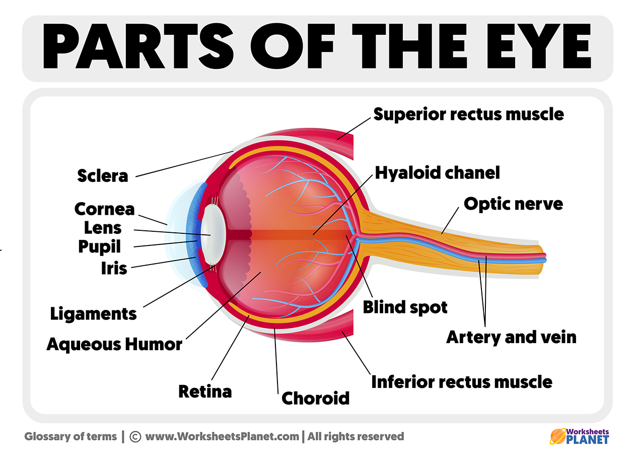
The human eye has a complex structure that can be divided into three parts:
- The outer layer comprises the conjunctiva, sclera, and cornea.
- The middle layer is composed of the lens, the ciliary body, and the iris.
- The inner layer, in which the pupil, retina, and vitreous humor are found.
Conjunctiva
The conjunctiva is a transparent mucous membrane that protects the inside of the eyelids and the sclera. It is composed of two continuous sections that prevent foreign elements from accessing the back of the eye:
- The bulbar conjunctiva covers the front part of the sclera.
- The palpebral conjunctiva veils the inner part of the eyelids.
The sclera: The white part of the eye
The sclera or sclera, informally known as the white part of the eye, is the structure that safeguards the organs of the visual system. Composed of collagen fibers, it supports the extrinsic muscles that facilitate eye movements.
The cornea
Located in the front part of the eye, on the iris, cornea is a convex spherical cap responsible for refracting light and transferring it to the retina.
The crystalline
The lens is the eye’s lens, whose main function is to focus on objects regardless of distance. To achieve this, it uses the accommodation process, which can modify its curvature, thickness, and refractive power depending on the circumstances with the help of the ciliary muscles.
The ciliary body
The ciliary body is a muscle located behind the iris and is responsible for changing the shape of the lens to focus vision. In addition, it produces aqueous humor inside the eye.
The iris
In addition to determining the color of the eyes, the iris allows the pupil to change its size to regulate the amount of light that enters due to the small muscles that form it, called orbicularis.
The pupil: the opening of the eye
The pupil is a hole in the iris through which light enters the inner part of the eye. By changing the size, it regulates the amount of luminosity that enters.vitreous humor. Transparent in color and gelatinous in texture, the vitreous humor is found inside the eyeball to allow the retina to remain in place and the eye to retain its spherical shape.
The retina
The retina transforms the light that enters the eye into electrical stimuli through the photoreceptors, which can be of two types:
- Cones, which allow us to distinguish colors;
- Rods, who appreciate black and white.
The macula
The macula is located in the retina’s center and is directly responsible for seeing details and capturing movement.
Peripheral retina
The peripheral retina allows us to identify the shape and silhouette of objects outside our direct line of sight. It detects, therefore, what we have around us.
The Optic nerve
The optic nerve, which is made up of thousands of fibers, sends visual signals to the brain.
